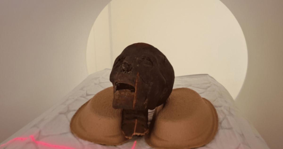

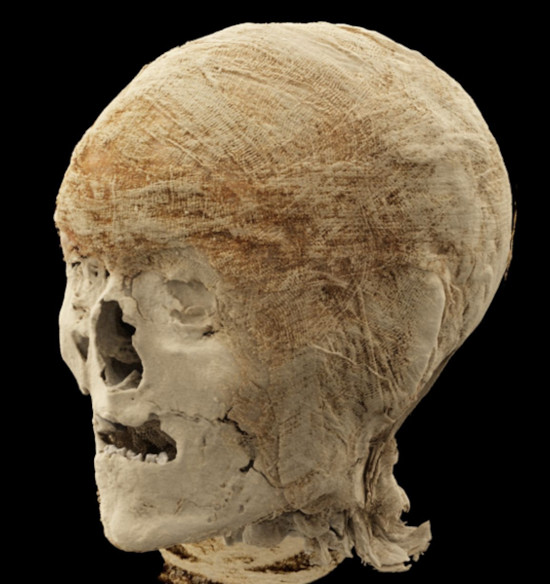
A mummified head examined at Semmelweis University using photon-counting CT technology. (Medical Imaging Center (OKK), MNMKK Semmelweis Museum of Medical History/ Semmelweis University)
Conducted during the night to accommodate standard clinical practices, the examinations focused on a collection of mummies that have been part of the museum's collection since its founding. Although these remains had previously undergone various imaging and multidisciplinary tests, including conventional CT scans, earlier technological constraints prevented a truly detailed assessment. Now, the new imaging technology is offering a non-destructive way to analyze the complex, multilayered materials of the mummified human remains. According to Dr. Ibolyka Dudás, Chief Clinical Physician at the Department of Radiology and Head of the working group for post-mortem imaging:
"The aim of the examinations is to obtain as accurate a picture as possible of the internal structure of the remains, any abnormalities, and the preservation techniques used."
- Scans of 2,000-Year-Old Egyptian Mummies Reveal Priestly Ailments
- Three 2000-year-old Egyptian Mummy Faces Reconstructed!
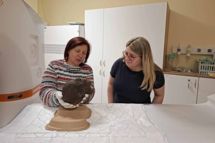
Krisztina Scheffer (MNMKK Semmelweis Museum of Medical History) and Dr. Ibolyka Dudás (OKK) holding the mummified head of an Egyptian woman. (Medical Imaging Center (OKK), MNMKK Semmelweis Museum of Medical History)
Unveiling the Past with High-Resolution Imaging
Among the specimens examined were two mummified heads, and the recent high-resolution CT scans have allowed researchers to conduct a far more detailed examination of the teeth and skull sutures than was previously possible. This enhanced visibility could lead to a more accurate age determination for the individuals and lay the groundwork for future high-precision, detailed 3D reconstructions, as well as potential facial reconstructions of the skulls. Such reconstructions have become increasingly popular in archaeological studies, offering a tangible connection to the people of the past.
In addition to the heads, the researchers examined a left lower limb that had previously defied a definitive diagnosis. Based on the new images, several possible interpretations have emerged. The current examinations indicate that the individual may have suffered from osteoporosis; however, the exact cause—whether due to age-related factors or a pathological process—requires further, detailed analysis. A second lower left limb also yielded new findings: the remains likely belong to a young individual, and this is the first time such detailed imaging data has become available for this particular find.
- Mummies Scanned to Unravel the Beautiful Mysteries Bound Inside
- 5 Discoveries That Changed Our Understanding of Mummification
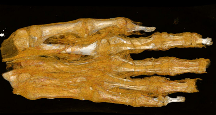
The mummified hand included in the study can offer valuable information about the individual's sex and age. (Medical Imaging Center (OKK), MNMKK Semmelweis Museum of Medical History/Semmelweis University)
Reinterpreting Ancient Remains
Another fascinating discovery involved a set of remains that had previously been interpreted merely as a mummy bundle. When the find first arrived at the museum, without the benefit of imaging tests, it was initially identified as a human head and later, possibly, as a bird mummy. However, a previous CT scan clearly showed that the find is, in fact, an adult foot. The current imaging analysis aims to determine the extent to which the textile remnants can provide insight into the mummification technique, the age of the mummified individual, and any illnesses the individual may have had. The current images clearly reveal the different layers of the bandage and show their different structural characteristics, which may lay the groundwork for further historical and technological investigations. The remains were presumably part of a complete mummy, but the reason and time of the dissection are currently unknown.
The analysis of a mummified hand included in the study is also expected to offer valuable information. Based on the size, development, and morphological characteristics of the bones, researchers aim to determine whether the remains belonged to a child or an adult. They may also be able to estimate the individual's sex and age. Radiocarbon dating on some of the specimens has already revealed that the oldest remains can be dated to between 401 and 259 BCE, making them more than 2,300 years old. Six specimens were subjected to C14 dating, though only three yielded measurable results.
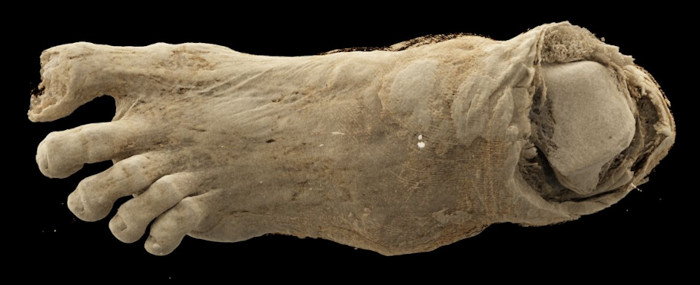
Detailed image of a mummified foot produced with the news scanning technique. (Medical Imaging Center (OKK), MNMKK Semmelweis Museum of Medical History/Semmelweis University)
A New Era for Mummy Research
The application of cutting-edge technology is opening new doors in the field of mummy research. Chief Museologist Krisztina Scheffer emphasized that while the remains had previously been examined by a research team, the current images provide a more detailed view than ever before and are expected to yield new, scientifically valid findings regarding the remains that have been preserved in the collection for decades. A detailed evaluation of the images is currently underway, and researchers expect the data analysis to provide new insights into the mummies' lives and state of health, as well as the mummification process itself.
The photon-counting CT scanner at the heart of this research represents a significant leap forward in medical imaging technology. Unlike conventional CT scanners, photon-counting detectors capture more detailed information about the energy of individual X-ray photons, resulting in sharper images with greater contrast and reduced noise. This makes them particularly well-suited for examining the dense, complex, and multilayered structures found in ancient mummies. As Scheffer noted, "Based on the results so far, it is evident that modern imaging technology opens up new perspectives in mummy research. It can reveal information hidden in finds that are thousands of years old without damaging them." This non-invasive approach is crucial for preserving these invaluable artifacts while still extracting the wealth of historical and medical data they hold, and the ongoing analysis at Semmelweis University is set to contribute meaningfully to our understanding of ancient Egyptian health and disease.
Top image: A mummified head going through a CT scanner. Source: Medical Imaging Center (OKK), MNMKK Semmelweis Museum of Medical History/ Semmelweis University
By Gary Manners
References
Szvák, E. 2026. Egyptian mummy remains examined at Semmelweis University. Semmelweis University. Available at: https://semmelweis.hu/english/2026/04/egyptian-mummy-remains-examined-at-semmelweis-university/